
Unit 3 Introduction
This unit introduces some of the major techniques with which scientists gain understanding of the structure and function of the human brain. What kinds of experimentation help us better understand how neurons communicate and how different parts of the brain work together? Some methods can be used to directly alter brain activity in a living person, while others merely record the brain’s activity or create a map of where that activity is located. Students will learn which methods can be used without damaging or permanently impacting the subjects, and which are best for clinical uses. Students will also discuss ethical concerns that exist surrounding the development and use of these technologies. Lastly, this unit covers current methods in animal systems that are revealing new insights into the human brain.
What's In This Unit?
- Lesson 1: Non-Invasive Methods in Humans
- Lesson 2: Invasive Methods in Humans
- Lesson 3: Ethical Issues with Neuroscience Methods
- Lesson 4: Methods in Animal Research
Terms & Definitions: Unit 3
- MRI/fMRI – Magnetic Resonance Imaging (MRI) uses magnetic fields and radio waves to obtain a picture of the brain (or other organs/tissue) inside the body. Functional Magnetic Resonance Imaging (fMRI) uses this same technology, with some additional adjustments, to look at blood flow in the brain over time, which indicates real-time brain activity, often while the subject is engaged in a cognitive task.
- PET – Positron Emission Tomography (PET) uses a radioactive tracer (which is injected or swallowed) to measure brain function.
- CT – Computed Tomography (CT) is a type of 3-D X-ray that can be used to image the brain.
- EEG – An Electroencephalogram (EEG) uses multiple electrodes placed on the surface of the skull to measure the electrical activity in the brain.
- Temporal resolution - How fast a brain imaging method can gather data of the brain’s activity. Methods with high temporal resolution can give detailed information about rapidly changing patterns of activity in the brain.
- Spatial resolution - How small an area of the brain an imaging method can gather data from. Methods with high spatial resolution can give detailed information about the structure of the brain or where in the brain certain activity is. Much like the number of pixels on a screen, the more chunks the brain is divided into, the sharper and crisper the image will be.
Lesson 1: Non-Invasive Neuroscience Methods
Objective: Students will be able to explain the advantages and disadvantages of the most common non-invasive methods currently used in neuroscience and their potential for treating brain disorders and mental health conditions.
ENGAGE/HOOK: A First Look at Imaging (5 min)
In order to activate students’ observational skills, ask them to analyze the images below of different technical brain imaging methods.
Explain that there are many different ways to get a visual representation of what is going on inside the head. Each method has strengths and weaknesses, and because of this, certain methods are typically used in certain settings.
Show students the images below and ask the the questions that follow:
Image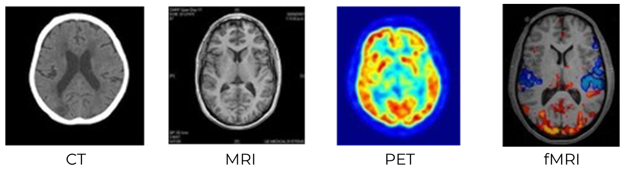
- Ask students to answer the following questions independently:
- Which images are the sharpest? Which are the fuzziest?
- Which images have intensity or color scales? What do you think the colors or intensities might signify in each image?
- Compare the CT/MRI images with the PET/fMRI. What differences and similarities do they see between these groups?
Things to note that will help you during student discussion:
- If students point out that MRI and fMRI look sharper than CT and PET, explain that the date of development of each technology correlates with the increasing image resolution. CT was introduced in the early 1970s, PET and MRI in the 1980s, and fMRI in the 1990s. Most present-day research in cognitive neuroscience uses fMRI when looking at brain activity for this very reason.
- If students point out that CT doesn’t show the difference between the white matter and gray matter within the brain, you can explain that this is why CT is not commonly used for neuroscience experiments but is often used by doctors and hospitals to see the location of tumors or areas of damage within the brain. Refer to Unit 1, Lesson 5 for more on gray and white matter.
EXPLORE: Mapping the Brain (20 min)
Use the Mapping the Brain interactive activity from NOVA scienceNOW to help students explore several non-invasive imaging techniques. Guide them through the activity using the steps below.
- Provide students with the Mapping the Brain worksheet.
- Project the online simulation to the whole class to demonstrate how to navigate the brain anatomy in the interactive online module. Abbreviated directions from the worksheet on how to navigate the brain in the online module can be seen below:
- Select one cross-section of the brain to view using the three buttons at the top (coronal, axial, or sagittal).
- Coronal cross sections divide the front of the head from the back.
- Sagittal cross sections divide the left side of the head from the right.
- Axial cross sections divide the top of the head from the bottom.
- The slider at the bottom allows you to scroll through the brain. Notice the yellow-green line through the head on the left side of your screen moves along with you as you scroll, showing you where you are within the head.
- The menu on the right shows all the different brain regions that you can select.
- The slider at the right changes the color transparency of the selected brain region.
- Select one cross-section of the brain to view using the three buttons at the top (coronal, axial, or sagittal).
- Students will select one of the brain regions in the menu on the right-hand side of the screen and answer questions 1-3 in the worksheet. If time allows, you can have students choose a second brain region and answer questions 1-3 again.
- Once students complete the worksheet, lead a discussion reflection:
- How does imaging technology help scientists research the brain?
- Why might scientists use more than one imaging technique when conducting their research?
EXPLAIN: Advantages and Disadvantages of Non-Invasive Techniques
The following information is teacher-facing and can be utilized to teach students new information in whatever format works best for you and your students.
Key points:
- Non-invasive methods can be used to study the structure and function of the brain, as well as to modify brain activity.
- Different methods measure different indicators of brain activity, such as electrical signals (EEG), blood oxygen levels (fMRI), or radioactive tracers (PET).
- The accuracy and resolution of timing and spatial location of brain activity are two major characteristics to consider when comparing the benefits and disadvantages of different methods.
In Unit 1, Lesson 4, we saw how lesion studies were used historically to learn about the roles and functions of specific brain regions. However, the accident or event that caused the lesion in question may have other impacts in other regions of the brain, so it is hard to be certain that a particular symptom is purely due to a particular lesion or brain region. Modern techniques offer a more sophisticated way to look at healthy brains, to see what their structure looks like and which regions are active during particular tasks or events.
Techniques for Studying Brain Structure
- The most common techniques for getting highly detailed images of the brain are computed tomography (CT) and magnetic resonance imaging (MRI). A CT scan combines X-ray technology with computer processing to create detailed cross-sectional images of the body. MRI uses a strong magnetic field and radio waves to generate detailed images of the body's internal structures. MRI provides valuable information about soft tissues, organs, and structures that may not be as clearly visible with other imaging methods. CT scans, however, are much faster than MRIs (10 minutes vs. up to an hour) and are preferred for emergency situations requiring immediate treatment, like a stroke.
Diffusion tensor imaging (DTI) is a specialized MRI that is used to visualize and analyze the structural connectivity and organization of the brain's neural pathways (made up of white matter), as seen in the image below. DTI uses the movement of water molecules within brain tissue to infer the orientation, direction (meaning which way action potentials will move down these axons), and overall organization of these bundles of white matter.
Image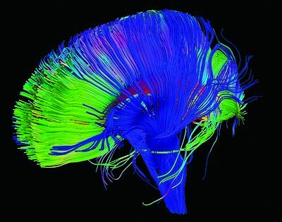
DTI imaging creates detailed and beautiful color coded images of white matter bundles. Image credit: NICHD/P. Basser
Techniques for Studying Brain Activity or Function
Other types of non-invasive techniques can measure neural activity and other kinds of brain changes at the very same time that people are performing mental tasks or psychology experiments. These methods allow us to pinpoint which areas of the brain seem to be involved in specific thought processes.
Electroencephalography (EEG) is a non-invasive diagnostic test that measures and records the electrical activity of the brain. EEG is used to diagnose and monitor brain activity associated with epilepsy, sleep disorders, and other neurological conditions. This method can also be used to look at neural activity relating to a task or stimulus in an experiment. EEG can measure brain activity almost instantaneously, giving high quality information about when that activity occurs (temporal resolution), but is not very accurate about where it occurs (spatial resolution).
Image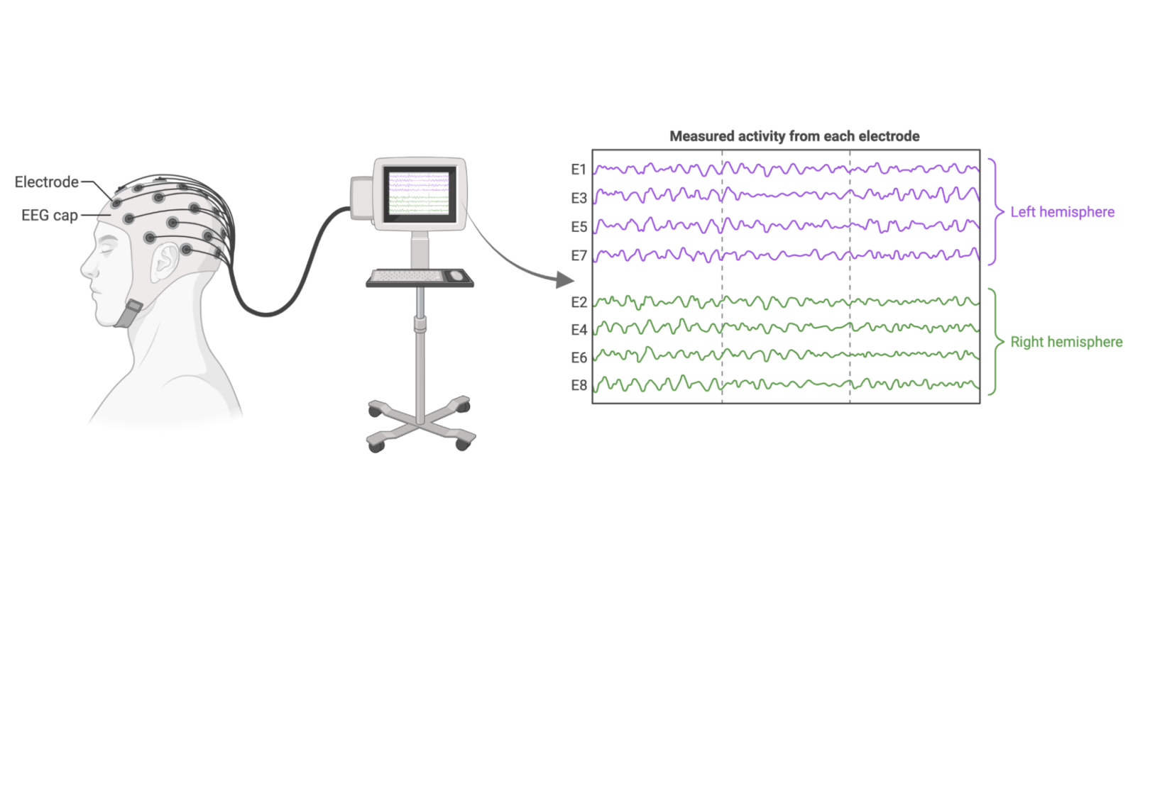
EEG directly records the electrical activity of neurons at the surface of the brain. Data from each electrode in the EEG cap is plotted on a graph in order to track brain waves relating to sleep patterns, seizures, or other changes in electrical activity. Image credit: BioRender.com
- Functional Magnetic Resonance Imaging (fMRI) relies on the fact that when a particular region of the brain becomes more active, there is an increase in blood flow and oxygenation to that specific area. By comparing the images taken during different tasks or conditions, we can identify which regions of the brain are activated or deactivated. However, fMRI is a slow technique. This means that it has high spatial resolution and takes crisp detailed pictures of the brain structure but since it has low temporal resolution, it can only take a new picture of the brain’s activity every 2 seconds (during which time the brain can do many different things!). It is also important to remember that there are many inferential and statistical steps in between the raw fMRI data and a pretty picture of a region that “lights up” with neural activity during a task! Many types of computer processing are required to create the final image.
- Positron Emission Tomography (PET) is used to study brain function by measuring the metabolic activity and blood flow in different regions of the brain. After a person is injected with a radioactively labeled tracer molecule similar to glucose, oxygen, or a neurotransmitter, a PET scan can follow the uptake and use of the tracer by different brain regions. PET scans have high accuracy in measuring biological function, but their spatial and temporal resolution are limited by the properties of the detector.
Magnetoencephalography (MEG) measures the magnetic fields generated by the electrical activity of the brain; the image below shows an example of brain activity while viewing a visual pattern. MEG has high temporal resolution, but its spatial resolution is limited to areas close to the scalp.
Image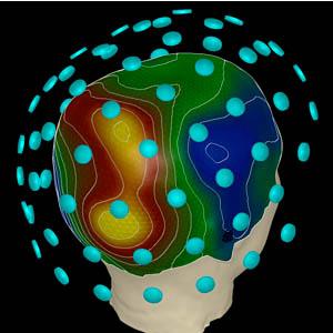
MEG imaging can be used to create a heatmap of where brain activity is occurring. Image credit: Los Alamos National Laboratory
Modifying Brain Activity
Beyond characterizing brain structure and function, newer non-invasive technologies such as transcranial magnetic stimulation (TMS) or transcranial direct current stimulation (tDCS) allow researchers to alter neural activity in a region of the brain by applying electrical or magnetic stimulation. This can increase or decrease activity in neural circuits in the region where the instrument is applied. These techniques are often compared to electroconvulsive therapy (ECT) because they change brain activity, but they do so in a more specific and more targeted fashion. These techniques are being studied for both therapeutic and enhancement purposes; they are addressed in more detail in Unit 7, Lesson 1.
Additional Resources
- Overview of current methods
- This website gives a nice overview of various methods of neuroimaging, with links to other sources. (McGill University)
- The image galleries on this site include collections of images from brain researchers that show different techniques used in current research and their applications to understanding the brain and brain health. (The Franklin Institute)
- This interview with neuroscientist Wei-Chung Allen Lee describes why and how researchers aim to understand the connectivity of the brain. (Harvard Medical School)
- Neuroimaging can get very technical very quickly, but here are some videos that explain the basics of the methods:
- EEG: This video gives a good overview of electroencephalography techniques. (2-Minute Neuroscience)
- fMRI: This video with Alan Alda is engaging and very simply explains how functional magnetic resonance imaging works and what it’s like to be in the scanner. (PBS/Brains On Trial)
- MRI: A brief video on how general magnetic resonance imaging works. (National Institute of Biomedical Imaging and Bioengineering)
- PET: A brief video on the methods of positron emission tomography. (National Institute of Biomedical Imaging and Bioengineering)
- MEG: This video gives a good overview of current magnetoencephalography methods and how it is used in conjunction with other technologies. (Alt Shift X/ARC Centre of Excellence in Cognition and its Disorders)
- Comparisons and limitations
- This article compares the differences between CT and MRI. (Healthline)
- This neuroscience blog post directly compares the strengths and weaknesses of PET and fMRI. (Brainy Behavior)
- This article is a helpful summary of how fMRI data can be misinterpreted and how other complementary techniques can offer a more reliable understanding of the brain. (Vox)
ELABORATE: Choosing the Right Methods (20 min)
Have students complete the following activity considering when different types of imaging techniques are appropriate. Students can refer to the Non-Invasive Imaging Methods student guide for reference.
- Have students read the 4 different scenarios below.
- Scenario #1: A baseball pitcher gets hit in the head by a line drive. Which method(s) would you use to determine what injuries he may have sustained? (CT for rapid screening for fractures, MRI to assess soft tissue damage)
- Scenario #2: You want to characterize brain health during aging by measuring sleep quality and performance on memory tasks. Which method(s) would you use to predict brain health using these measures? (EEG for sleep quality, fMRI for cognitive function, MRI for brain structure analysis)
- Scenario #3: You want to determine how fast dopamine is produced and neurons are activated after a person with Parkinson’s disease receives a new medication. Which method(s) would you use to look at the brain’s response? (PET for dopamine production, fMRI for neuronal activity)
- Scenario #4: You want to study how brain activity across a network of brain regions differs between veterans who developed post-traumatic stress disorder (PTSD) and those who did not, following a brain injury in combat. Which method(s) would you use to look at brain network activity? (MEG or EEG for brain activity, MRI for identifying brain regions)
- Ask students to consider what type of imaging method is best for each different situation. Students may use this reference sheet to recall the different imaging techniques.
- For each scenario ask students to answer the following questions:
- What type of imaging technique to use. Include multiple methods if appropriate.
- Information about how the methodologies work and why they are best suited to this scenario.
The pros and cons of different methods for this scenario.
Lesson 2: Invasive Methods
Objective: Students will be able to explain the value and applications of invasive neurosurgical techniques in learning about the brain and in treating brain disorders.
ENGAGE/HOOK: Why Use Invasive Methods? (10 min)
To introduce the idea of invasive methods guide students through the following intro activity:
- Show students this video of a deep brain stimulation surgery performed at the Northwestern University Department of Neurosurgery. Note that the video shows footage of live surgery, but is not particularly graphic.
- As they watch, ask students to answer the following questions:
- Why do you think the patient and her medical team opted to try this surgery?
- What non-invasive methods were used as part of the surgery prep? What information did they provide for the surgery team?
- How was the patient monitored during the surgery?
- How do you think they determined the surgery was successful?
EXPLORE: Early Development of Invasive Methods (30 min)
Explain the background information below to students to frame the following activity:
Wilder Penfield (1891-1976) was a Canadian neurosurgeon and pioneering researcher in the field of epilepsy and neurology. Penfield’s work using invasive neurosurgical techniques had a major impact on our modern understanding of the brain. He developed a surgical procedure called the "Montreal Procedure" for the treatment of epilepsy. During this procedure, he could electrically stimulate specific areas of the brain in conscious patients undergoing neurosurgery. This allowed him to create detailed maps of the brain and identify functional areas responsible for various sensory, motor, and cognitive functions.
- Have students read this short article from Quartz about Wilder Penfield and his work. Then watch this video about Penfield’s famous experiment.
- Show them the image of the sensory homunculus below. The video above shows the electrical stimulation of a patient’s brain stimulating particular memories, but Penfield also used this technique on the sensory areas of the cerebral cortex to map out this region of the brain.
- Image
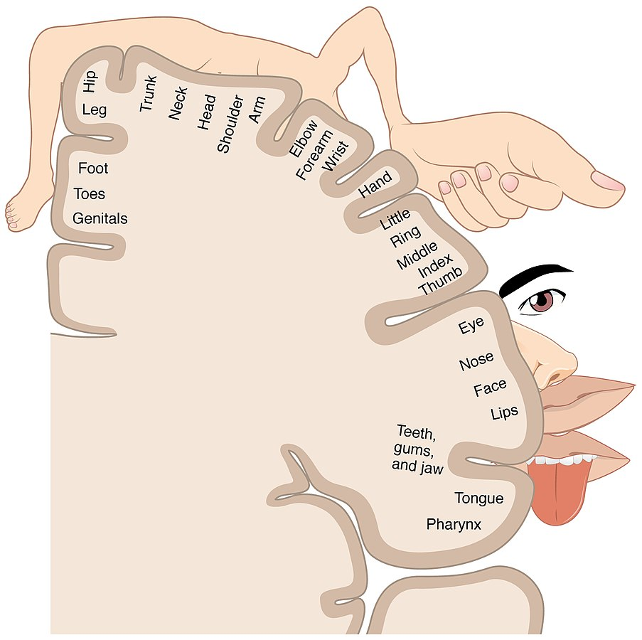
Many regions of the cerebral cortex, including the part responsible for processing touch and body sensation, contain a spatial map of the sensory environment (in this case, the skin). Image credit: OpenStax, licensed under the Creative Commons Attribution 3.0 Unported license.
- Ask students to independently answer the questions below and then discuss with table partners or as a class:
- What does the sensory homunculus diagram represent?
- What is the “Montreal Procedure,” and how did the technique allow Wilder Penfield to create the diagram?
- How were Penfield’s techniques similar or different to what you observed in the video of modern deep brain stimulation surgery?
EXPLAIN: Invasive Methods in Humans
The following information is teacher-facing and can be utilized to teach students new information in whatever format works best for you and your students.
Key Points:
- Invasive techniques of studying the brain yield more precise information about the stimuli, timing, and location of brain activity than non-invasive techniques.
- Devices implanted directly into the brain can modulate the brain’s electrical activity to treat some brain disorders.
- Signals recorded from the brain can be interpreted by computers to control external devices or prosthetics.
In order to gain insight into how the brain works, scientists from long ago used to need direct access to the brain so they could record the activity from neurons or stimulate those cells with electricity—like Penfield’s experiments. As described in Lesson 1, new technologies have made it possible to both record from neurons and stimulate without drilling a hole in the skull, but invasive methods can still be helpful, especially for direct medical intervention.
Treating Brain Disorders
Deep Brain Stimulation (DBS) involves the implantation of a device that can deliver electrical impulses to these targeted areas to modulate and regulate the electrical activity in the brain, especially for managing symptoms of movement disorders such as Parkinson’s disease, tremor, and epilepsy. Electrodes are surgically inserted through holes in the skull into the targeted brain regions. The electrodes are then connected to the stimulator, which is implanted in the body. After surgery, the device is programmed for optimal electrical stimulation to manage the patient’s symptoms.
Connecting to External Devices
A Brain-Computer Interface (BCI) is a technology that allows direct communication between the human brain and an external computer or prosthetic device without having to be relayed through the muscles or limbs. In general, a BCI records brain activity signals, then interprets and translates them into commands or actions that can be used to control the external device. Surgically implanted BCIs are typically used for users with serious disorders or injuries. There are also non-invasive wearable BCIs, but these are more commonly used for nonmedical purposes. Most BCIs are currently limited to experimental applications. Reference Unit 7, Lesson 2 to see more on these state-of-the-art methodologies.
Direct Measurement of Brain Activity
Intracranial recording is a technique used to directly measure electrical activity within the brain. It involves placing electrodes directly onto or inside the brain tissue to record and analyze neural signals. Intracranial recording provides powerful data because of its precision and high spatial and temporal resolution. For medical purposes, this technique is typically used in patients with epilepsy or other conditions that require precise mapping of brain activity. However, once the electrodes have been placed they can also be used for nonmedical research purposes—importantly, with the patient’s consent—to record other types of information that are processed in those brain areas.
Additional Resources
- Treating Brain Disorders
- This website gives a good overview of Deep Brain Stimulation and the surgical process. (Johns Hopkins Medicine)
- Connecting to External Devices
- This video shows how a woman with tetraplegia (paralyzed from the neck down) was able to learn to control a robotic arm using a brain-computer interface. (Brown University)
- This 2022 report reviews the current technology of brain-computer interfaces as well as their opportunities and challenges. (U.S. Government Accountability Office)
- Direct Measurement of Brain Activity
- This 2022 article describes the process of intracranial recording and ethical issues around informed consent. (Science Magazine)
- In a 2022 study, scientists performed intracranial recording on epilepsy patients with electrodes placed in their auditory cortex and had them listen to different types of sounds. After analyzing brain activity, the scientists discovered a population of neurons that responds specifically to song! (MIT Technology Review)
ELABORATE: Mapping Your Brain (45 min)
Explain the background information below to students to frame the following activity:
In this lesson we have seen how invasive techniques of studying the brain can provide critical insights into how, where, and when the brain processes different types of information, and how these insights can be used for new types of therapy for brain disorders and injuries. Now try mapping your own brain! We will model the results of using invasive measurement technologies by using a technique called the two-point discrimination task.
Have students create their own sensory homunculus using this Brain Maps handout from BrainFacts.org as a guide. The basic steps are broken down below:
- Provide students with a brief background lecture using points from the Brain Maps handout (pgs 1-3). These slides may also help. Introduce the activity to students: “Today you will explore brain mapping in a very personal way. Each of you will create your own individual homunculus.”
- Students create the two-point discrimination tool. Directions are on pages 3-4 of the handout.
- Students measure the two-point discrimination threshold at various places on the body and record results.
Students create an individual sensory homunculus using their own data.
Image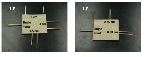
Students will create small two-point discrimination tools with classroom items such as toothpicks, tape, glue, and thick paper. Image credit: BrainFacts.org
Lesson 3: Ethical Issues with Neuroscience Methods
Objective: Students will be able to explain a few common ethical concerns relating to the most common methods of cognitive neuroscience.
ENGAGE/HOOK: Who’s Included in Research? (15 min)
Introduce the lesson by explaining the background information below and guiding them through the following activity:
This lesson will touch on topics of diversity, inclusion, and other ethical considerations in the field of neuroscience.
- To get students thinking have them discuss:
- If the brain is the same organ, why does diversity in neuroscience subjects matter?
- Think about what you know about human physiology, genetics, and the role of nature vs. nurture in shaping the brain. Why is it important to include:
- Diversity in sex and gender?
- Diversity in genetic ancestry?
- Diversity in geographic location?
- Show students this video of researchers hoping to recruit people in rural India to participate in brain research. As they watch ask them to answer:
- Why are the researchers using a simple EEG device?
- What are some of the technical challenges faced by the researcher?
- What are some of the values and concerns expressed by people in the village?
- Explain to students: To date, the majority of neuroscience research has been conducted in relatively few countries—the U.S., Europe, Australia, China, and Japan. Neuroscience research is better and creates more accurate science when diverse people are included, but as we will see in this lesson, there are social and ethical issues that must be addressed.
- If students are familiar with examples of historical discrimination and abuse in biomedical research of African American communities in the U.S. (e.g., the cases of Henrietta Lacks and the Tuskegee Syphilis Study), extend the discussion to connect with the impacts of this cultural distrust. This NPR article discusses an example of research initiatives aiming to overcome that troubling legacy (not specific to neuroscience).
EXPLORE: Equity and Access to EEG (30 min)
Guide students through the analysis of the following case study about a patient with chronic migraines who was almost denied a medically necessary EEG due to her hair texture and style.
- Have students read this article from KFF Health News about the patient’s challenges with getting an EEG.
- After reading ask students to independently answer the questions below:
- What were the instructions given to Sadé by the medical facility? Why are these instructions “anti-Black”?
- How do you think these instructions made Sadé, and other Black and Brown patients feel?
- Does the article mention any methods that get around these hair restrictions? What are they?
- If a facility does not have ways around the restrictions, how could they more inclusively communicate these barriers to patients with thick hair, braids, or extensions/wigs?
- Lead a whole class discussion after students have had a chance to independently reflect.
EXPLAIN: Ethical Considerations of Common Methods
The following information is teacher-facing and can be utilized to teach students new information in whatever format works best for you and your students.
Key Points:
- Like any rapidly evolving technology, brain imaging raises several important ethical considerations.
- Brain imaging devices should be designed to make sure everyone has equal access to these techniques and research data are more representative of the population.
- Even scientists can fall victim to logical fallacies in interpreting brain imaging data based on their own preconceptions.
- The use of AI for analyzing fMRI data to decode the specific meaning of neural activity patterns is raising new concerns about privacy and consent.
Implicit Bias in EEG Research
If you, personally, haven’t encountered a particular issue before, then you may not factor in that issue when designing new technology. One example is that electrodes for EEG technology were primarily designed by people of Caucasian descent who tended to have thin, often straight, hair. As a result, existing EEG electrodes can be difficult to apply to people with curlier, thicker, and more highly textured hair including many Black and multiracial people. These barriers have produced an overrepresentation of people of Caucasian descent in EEG research data, skewing research findings. Today, scientists are redesigning EEG electrodes to work well with thick textured hair and can be tucked under braids to fit snugly against the scalp. This work is being led by many scientists and engineers who themselves are Black and have hair of this type, in order to correct the problems in the field. Similar biases are embedded in other methods as well, affecting not just inclusion in research but also the accuracy of medical diagnostics.
Drawing the Wrong Conclusions
Many neuroscience research studies are designed to assign the subject a certain task or stimulus and then observe imaging data of the resulting brain activation. However, sometimes studies start by looking at brain imaging data and work backwards to draw conclusions about how someone is feeling or what they are experiencing based solely upon their brain activity or structure. The problem with this is that each region of the brain can be involved in many different tasks and it is impossible to know exactly what a particular activity pattern means from imaging alone, but scientists and journalists can often make this mistake due to preconceived expectations.
Decoding Your Thoughts
With artificial intelligence and machine learning, more sophisticated analysis of fMRI imaging data is making it possible to decode what someone is thinking. An AI decoder can be trained to process a person’s brain activity patterns that are associated with known mental states or thoughts. When the trained decoder is applied to new fMRI data, it maps the new data to the training patterns to predict what the person is thinking. Such neural decoding experiments raise ethical concerns about consent and how to protect the privacy of your thoughts—especially when it comes to issues of personal identity such as health, gender, or sexuality—as technology continues to advance. (See more details on AI and future technologies in Unit 7, Lesson 4.)
Additional Resources
- Implicit Bias in EEG
- This article describes the research into developing EEG electrodes that work with more types of hairand includes interviews with young Black scientists. (Massive Science)
- This article explains the implicit biases in various neuroimaging technologies and methods of analysis. (Nautilus)
- Students may be familiar with similar technical issues that came to light during the Covid-19 pandemic with pulse oximeters that didn’t work as well on people with darker skin. This article highlights researchers trying to fix the problem. (STAT News)
- Drawing the Wrong Conclusions
- This interview with fMRI expert Russ Poldrack highlights fallacies common in interpreting neuroimaging research and the idea that scientists and companies are trying to improve brain imaging so as to be able to “read our minds.” (The Verge)
- This blog post written by graduate students in neuroscience explains the idea of “reverse inference,”with some key examples. (Knowing Neurons)
- Decoding Your Thoughts
- This article discusses how artificial intelligence can be used to analyze fMRI to try to predict behavior or “read minds,” and what ethical problems may result. (CNN)
- This article from NPR describes a recent study from the University of Texas at Austin where scientists used fMRI and AI to detect stories in people’s minds by decoding language patterns. (NPR)
ELABORATE: The Ethics of Brain Reading (30 min)
Explain the background information below to students to frame the following activity.
Although it is currently not possible to use any method of brain imaging to truly “read your mind” with reliable accuracy, this may become possible in the future. As discussed earlier in the lesson, we can record from human brains well enough to control a robotic arm, given enough training, but the same technology is not advanced enough to be able to decode or predict what a person is thinking. If brain imaging improves to the point that it IS possible to know what someone is thinking without them having to say or do anything, what are the ethical implications? One concern is about issues of privacy and how to keep one’s thoughts private. Another concern is how this information would be used—who would have access to the resources to read your mind without your permission?
Guide students through the following analysis using the steps below:
- Pose this question to students to get them thinking aloud:
Should neural decoding technology be used to read the minds of unconscious patients? - Have students read about this case study of a person who suffered damage to the cerebral cortex and was not visibly responsive, but could perform basic communication using a simple code.
- Give students this ethical decision making framework from the Northwest Association for Biomedical Research (teacher background guide) and research the facts and ethical issues associated with brain decoding in order to answer the question posed in #1.
Optional Example:
This article (and linked podcast) from NPR describes Martin Pistorius, a man from South Africa who developed locked-in syndrome. He had gone into a vegetative state at age 12 and remained so for years. When he started waking up, he was unable to move or communicate so none of his family members realized he had returned. Eventually he was able to use computer technology to communicate using his eyes.
Lesson 4: Methods in Animal Research
Objective: Students will be able to describe some common techniques used in animal experimental models that allow studies of the brain in cellular-level detail and with precise control of function.
ENGAGE/HOOK: New Methods, New Questions (10 min)
Introduce the lesson by explaining the background information below and guiding them through the following activity:
To begin thinking about animal methods in neuroscience we will analyze a study performed at Northwestern University that uses a recently developed technology in animal systems called optogenetics. This method allows researchers to turn on and off a targeted set of neurons using light to understand their function. (An article accompanying the video provides more background information on optogenetics and describes the details of the study.)
- Show students this video of research to study the brain circuits underlying social bonding.
- While they watch ask students to answer the following questions:
- How did the scientists program the mice’s behavior?
- What parts of the brain do you think the devices were connected to?
- What behaviors did you observe in the video when the devices were synchronized? Desynchronized?
- This experiment was the first time researchers used wireless devices for optogenetic control. Why was that advance important for investigating this particular research question?
- How do you think the results of this study might help us understand the human brain and behavior?
- Do you think this technology could be used in humans? Why or why not?
EXPLORE: Challenges of Human Methods (15 min)
Explain the background information below to students to frame the following activity.
Sometimes the study of certain brain activity is not physically or ethically possible in a human brain, and this is where animal methods come in!
Guide student groups through the following discussion in small groups:
- Ask small groups to list out the invasive and non-invasive neurological methods of study discussed in Lessons 1 and 2.
- Ask student groups to answer the following questions:
- Using what you know about the methods listed, what kind of experiments are possible to do in living humans and which are not?
- What kind of experiments might be easier or more ethical to conduct on animals instead of humans?
- Could you manipulate a person’s thoughts or behaviors and observe resulting changes in brain activity.
Answer: Yes. Intracranial recording allows us to do this in humans at a limited scale. - Could you manipulate cellular activity and observe the changes in thoughts or behaviors in humans?
Answer: No. Our current technologies don’t allow us to control the activity of individual cells in living humans. - Could you study the effects of a new drug that has only been tested in cell culture (cells growing in a dish in a lab) on human brain activity?
Answer: No. Drugs must be tested in animals before they can move to human trials. - Reflect on how our current methods for human research limit our understanding of brain activity and ethical considerations of studying human brains.
- Ask groups to share out their answers and find common themes in their responses.
EXPLAIN: Methods in Animal Research
The following information is teacher-facing and can be utilized to teach students new information in whatever format works best for you and your students.
Key Points:
- Experimental research on animals is a difficult ethical issue for some people, but many medical breakthroughs we enjoy today are due to well-designed and ethical animal research.
- Specific techniques in animals, such as Brainbow and optogenetics, are allowing scientists to map brain circuits and functions in unprecedented detail.
- Humanized animal models are laboratory animals that express human genes in order to learn more about human diseases and to test possible treatments.
Ethics of Animal Research
Going even further than the invasive methods used in humans, techniques developed for animal systems in recent years have given scientists the tools to map brain structure and manipulate function with unprecedented precision. These new methods are yielding new insight into human brain function and disorders. However, the use of animals in research is a controversial ethical issue. It is important to note that experiments using living animals are not performed lightly and go through a rigorous approval process. These experiments must be approved by a committee designed to minimize the number of animals used and the discomfort that these animals must experience. Veterinarians and animal care staff work to keep animals comfortable and happy, and scientists are required to be trained in animal handling and care. Many medical advances in neuroscience and beyond, including vaccines for polio and rabies, the development of some antibiotics, cancer treatments, and organ transplantation, have been developed thanks to the use of animals in research. These new treatments are not only used to improve human lives, but can be used in a veterinary setting to improve the health of animals as well.
Visualizing Connections
“Brainbow” is a technique used to visualize the complex connections within the brain. It uses a combination of genetic engineering and fluorescent proteins to color code individual cells in the brain. Researchers can then use advanced microscopy techniques to track and distinguish cells in regions of the brain, allowing them to study how neurons connect and interact with each other and change over time. It is most commonly used in mice, fruit flies, and zebrafish.
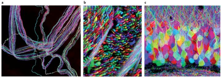
Brainbow images showing labeling of individual cells. Image credit: Lichtman & Sanes, 2008; licensed under the Creative Commons Attribution 3.0 Unported license.
Manipulating Function at a Cellular Level
Optogenetics combines genetic engineering and optics to control and manipulate the activity of specific neurons in living organisms, typically mice or other animals. It allows researchers to selectively activate or inhibit the activity of particular neurons with precise timing and spatial resolution, providing insights into the function and connectivity of neural circuits. Light-sensitive proteins are genetically introduced into target neurons to act like a switch—scientists can then use light to turn those neurons “on” and “off,” as shown in the illustration. By activating or inhibiting specific groups of neurons, scientists can observe the resulting changes in behavior, cognition, sensory processing, or other brain functions. This is similar to earlier studies on individuals with brain lesions, but more powerful as the precise regions of the brain turned off or on can be controlled at will by the experimenters.
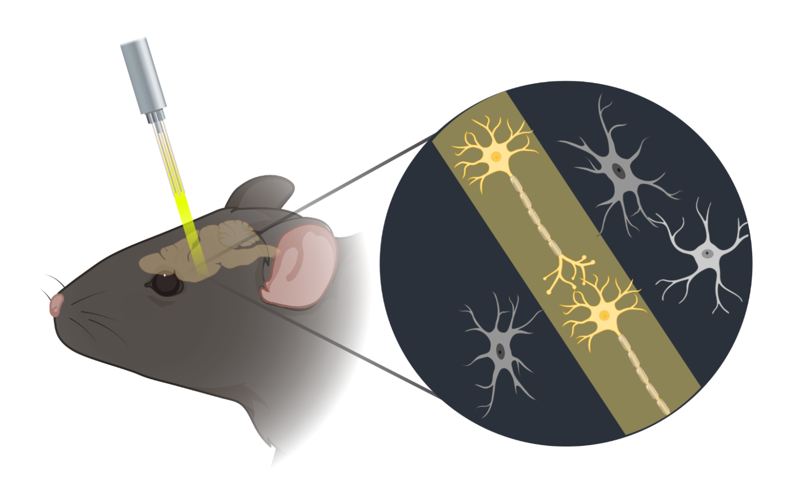
Optogenetics uses fiber optics implanted in to the head of the animal expressing light-sensitive proteins to turn the neurons containing those proteins on or off by changing their activity. Image credit: BioRender.com
Increasing Human Relevance
As discussed in Unit 1, Lesson 6, there is much we can learn from the brain structure and function of experimental animal models, but they’re still not human. How can scientists maintain the practical benefits of animal research while getting closer to understanding what actually happens in humans? “Humanized” animal models use genetic engineering and transplantation techniques to introduce human genes or cells into an animal model to mimic specific human traits or diseases for research purposes. For brain research, scientists create animals with human genes or cells to better model human development, physiology, and neurological disorders, or to get more informative data on the safety and efficacy of drugs and therapies in development before human testing.
Additional Resources
- Animal Research
- PBS Learning Media offers a short article and video to help grade 9-12 teachers and students explore the benefits of animal research and its ethical considerations.
- This article is a good overview of the protections in place to make scientific animal research as safe and ethical as possible. (The Conversation)
- Brainbow
- This video provides good visual analogies of how the Brainbow technique works and what information it reveals. (BrainFacts.org)
- This article includes examples of the beauty of Brainbow images. (Scientific American)
- This article discusses how Brainbow complements other non-invasive techniques in humans in efforts to map the entire “connectome,” or a full circuit diagram, of the brain. (BrainFacts.org)
- Optogenetics
- This video gives a good overview of the development of optogenetics and what it could be used for. (SciShow)
- This video interview with Ed Boyden at MIT, one of the pioneers of optogenetics, conveys his hopes of what the technology might be able to accomplish in humans someday. Note that in the nine years since this video was produced, gene therapy technology in humans has significantly advanced (using CRISPR gene editing, for example), removing one of the potential technical hurdles—though many more remain.
- Humanized animal experiments
- This article describes a study in which mice were genetically modified to express a human gene involved with Alzheimer’s disease to examine age-related changes in the brain and behavior. (National Institutes of Health)
- This article describes a study where Stanford researchers transplanted human neural cells into rat brains to investigate how the human cells could grow and function in the hybrid brain. More about experiments with brain organoids and their ethical considerations is covered in Unit 7, Lesson 3. (Fierce Biotech)
ELABORATE: Write a News Report
Explain the background information below to students to frame the following activity:
As we have learned from the technologies introduced in this lesson, technical advances in genetic engineering, electronics, and transplantation have opened up new avenues in animal research to understand the brain. However, these new techniques also raise many questions as to when and how they should be used. Communicating the potential benefits, challenges, and ethical issues to the public, and understanding public questions and concerns, is an essential part of responsible scientific research and innovation.
Ask students to script a short news segment about research on humanized animals in small groups using the steps below. Scripts should follow the “3 Cs of news writing”: be clear, concise, and correct.
- Break students into groups of 4
- Have groups select a humanized animal that has been genetically engineered or transplanted with human brain genes or cells to report on. They may choose one of the following articles as the focus of your script:
- Chinese scientists insert human brain gene into monkeys, spark ethical debate (NBC News)
- Supporting sources:
- Precision gene editing paves way for transgenic monkeys (Scientific American)
- Bioethical considerations for transgenic nonhuman primate models in neuroscience research (National Academies)
- Human neurons implanted in a rodent’s brain lead a rat to water—and make it drink (STAT News)
- Supporting sources:
- Human brain cells transplanted into rat brains hold promise for neuropsychiatric research (Stanford University)
- Are rats with human brain cells still just rats? (MIT Technology Review)
- Chinese scientists insert human brain gene into monkeys, spark ethical debate (NBC News)
- Ask students to write a script with the following roles:
- Anchor
- Scientific expert
- Ethicist
- Member of the public
- Students should use the following outline as a guide for their script:
- Anchor: Introduce the segment (~45 words)
- Scientific expert: Why was this research valuable? What was learned? (~200 words)
- Ethicist: What are some of the ethical concerns with this type of research? (~200 words)
- Member of the public: Do you think more research like this should be done? Why or why not? You may be creative about this person’s motivations and interest in the topic. (~150 words)
- Anchor: Closing/conclusion (~45 words)
Optional: Encourage students to film their script if time and resources allow.
Optional: Students can be asked to evaluate another group’s presentation for accuracy and helpful depiction of the ethical concerns.
For more information about the Neuroscience & Society Curriculum, please contact neuroscience@fi.edu.
Neuroscience & Society Curriculum
Launch Lesson • Unit 1: Neurons and Anatomy • Unit 2: Education and Development • Unit 3: Current Methods in Neuroscience • Unit 4: Mental Health and Mental Health Conditions • Unit 5: Drugs and Addiction • Unit 6: Law and Criminology • Unit 7: Future Technologies

This project was supported by funding from the National Institutes of Health Blueprint for Neuroscience Research under grant #R25DA033023 and additional funding from the Dana Foundation. Its content is solely the responsibility of the authors and does not necessarily represent the official views of NIH or the Dana Foundation.